Debridement is the process of removing dead, non-viable, poor-quality infected tissue and tissue residues, and foreign materials from the wound and its edges. Although the body normally tries to remove them naturally, if the amount is too much and especially in immunocompromised patients, this cleaning process is delayed and may not be successful. Wound debridement is a very important step in preparing the wound bed for healing. Preparation of the wound bed means the removal of necrotic, useless, non-healing tissues, control of exudate (discharge) and bacterial balance, and the creation of a suitable environment for the wound to heal. It is a scientific fact accepted by all wound healing experts that debridement is a necessary, useful, and effective procedure in wound care.
Aggressive surgical debridement is often required to remove dead and harmful tissues on and around the wound. The method of debridement is decided according to the condition of the wound and the needs of the patient. The knowledge and experience of the clinician dealing with the wound play an important role in deciding which method to use for each patient. The appropriate debridement method varies for each wound and its various stages. For this purpose, the wound should be carefully monitored during the process and the method to be used should be decided in multidisciplinary committees.
When surgical debridement is not feasible or appropriate, or for a quick and fine debridement without damaging healthy tissues, 'larval therapy' is a known, appropriate, and accepted scientific treatment method. Larval therapy is also called "maggot therapy" or "biosurgery". It is colloquially referred to as 'maggot therapy'.
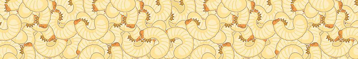
The larvae of Lucilia sericata (common green bottle fly) feed on dead tissue, tissue debris, and serous drainage material found in the wound. They physically break down and digest necrotic material and tissue debris. This process requires the physical activity of the larvae and proteolytic enzymatic digestion. The larvae secrete enzymes such as collagenase, trypsin, and chymotrypsin and eat the tissues after they have been broken down outside the body and converted into a semi-liquid state (digestion). Lucilia sericata larvae do not eat living, healthy human tissues. This selective behavior of the larvae is one of the most important advantages of larval debridement therapy, as it preserves healthy tissues necessary for healing. The number of bacteria in the wound is also reduced as bacteria as possible in the material, which is liquefied with the help of enzymes and digested and absorbed, will be digested, and absorbed, so it also has antibacterial effects. Larval secretions prevent the formation of biofilm and destroy the existing biofilm. Larval treatment can be performed in cases where rapid debridement of devitalized tissues that are thought to delay wound healing is required. Thus, the wound bed is prepared for wound care products or treatments (including surgical treatment) that will accelerate wound healing. While the larvae applied on the wound are initially very small, only 1-2 millimeters in length, they can reach up to 12 mm in length within 3-4 days during the wound cleaning process.

Larval treatment is an effective method of debridement and can be performed by any certified healthcare professional with adequate training, knowledge, and skills in wound care.
The application competence and application decision should be made by the doctor in charge of the wound care center, surgeon, wound care nurse, podiatrist, and most accurately by multidisciplinary boards related to wound care.
The patient is examined as a whole before the application of larvae. The type, cause, localization, and condition of the wound bed are carefully investigated and documented. Except for venous ulcers, wound healing is not possible if there is compression on the wound, the wound area and extremity should be free from compression. A careful vascular examination is performed, ischemia is documented, and surgical or endovascular revascularization is performed if possible and necessary. The patient and relatives are informed and informed consent is obtained.
Diabetic wounds, venous ulcers, arterial/ischemic wounds, mixed ulcers (arterial-venous), pressure sores, post-traumatic wounds, hematoma residues, necrotizing fasciitis (after surgical debridement), post-surgical pilonidal sinus, non-healing surgical wounds, wounds infected with 'Methicillin-resistant Staphylococcus aureus (MRSA)'. In addition to these, in other types of wounds where close follow-up of patients is carried out by multidisciplinary committees, the decision to treat larvae may be made by these committees with the condition of careful monitoring.
Contraindications
- Larvae should not be administered in patients with a bleeding tendency or risk and in cases where the vessels are very close to the veins and the veins are seen in an open wound.
- It should be used very carefully and under constant monitoring in areas close to sinus or fistula mouths.
- Use on dry necrotic eschar tissue is useless. The eschar should either be completely removed or opened in certain places, moistened, and softened to allow larvae to enter under this tissue.
- In patients using anticoagulant drugs, the efficacy of these drugs should be carefully monitored and if there are values above the limits, larvae treatment should not be performed due to the risk of bleeding.
- If it is to be used in wounds that open into a body cavity, where internal organs are exposed, great care should be taken, protective measures should be taken and close medical follow-up should be made.
- There may be a pain at the wound site (especially in ischemic wounds and in patients who already complained of pain before larval application). Pain control can be reorganized by reviewing the patient's previous analgesia. If the pain is severe, pain control should be ensured by removing the larvae from the wound and washing the wound.
- As with all other debridement methods, bleeding may occur due to damage to small capillaries, so it is important to check the wound every day.
- Local disinfectants, local anesthetics, and some hydrogels (especially those containing propylene glycol as a moisturizer and preservative) have negative effects on the survival and development of larvae, which in turn negatively affects treatment prospects. Therefore, residues of these substances should be carefully washed off before larval treatment and should not be used during larval treatment. Occlusive, very tight clothing, and bandages, wound care products are not used, compression bandages can be used with nonocclusive wound care products.
Application Options
There are mainly 2 application methods:
- Larval bags (BioBag): Larvae are placed in small, fine-textured, teabag-like pouches in which small pieces of foam are placed to protect the larvae. Depending on the type and size of the wound, different-sized pouches can be used. The larvae remain in these pouches during the whole treatment process. Depending on the condition of the wound and the progress of the treatment, BioBags are usually kept on the wound for 3 days (up to 4 days if necessary). If it is thought that there is not enough debridement after the application, new applications can be made.
- Free larval application: This is the only method used in our medical practice. The larvae are placed on the wound without being in a bag and allowed to move freely. Precautions should be taken to prevent them from overflowing out of the wound and prevent them from getting air. Depending on the location and size of the wound, these precautions are achieved by careful and diligent application of various wound care products. They also help to understand the depth of the wound and the actual localization of devitalized tissues. Their dense aggregation in certain areas and penetration deeper into the wound may indicate the need for deeper, more serious surgical debridement. Larvae are kept in special carrier containers before application. They should be kept at room temperature and used within 24 hours of production.
Before larvae application, the wound and its surroundings should be carefully cleaned by washing the wound thoroughly and removing all possible tissue debris and residues of wound care products. Once the skin is clean and dry, hydrocolloid dressings or nonallergenic transparent adhesives (best applied in negative pressure devices) are placed as close as possible to the wound edges to protect them from the harmful effects of excessive exudate and larval enzymes. Zinc pomades or barrier creams are applied to the surrounding skin that cannot be covered in this way. If there is maceration around the wound, these are also protected by applying zinc pomade liberally. The larvae are then placed on the wound carefully and without damaging the larvae.
If available, cover the wound with a special net-shaped cover that prevents the larvae from coming out by moistening it well. If not, several layers of sterile gauze are placed on the wound, again thoroughly moistened, and air permeability is ensured between the layers. The edges of the gauze (except on the wound) are glued in the same way on the transparent adhesive cover that was previously glued on the intact skin. Thus, the larvae are prevented from moving out of the wound. An absorbent pad that does not prevent air passage is placed on top. If this pad becomes very dirty with exudate, it should be changed at adequate intervals. In dry, non-draining wounds, they should be moistened frequently, not over-moisturizing and causing the larvae to suffocate. Finally, the area is wrapped with an air-permeable dressing. Film dressings or occlusive dressings should not be used, as the larvae will suffocate.
Pressure should be avoided on the larvae-treated area. While changing the soiled dressings on the wound, it should be observed that the larvae are alive and mobile. If the patient feels pain, discomfort, and restlessness, this should be carefully evaluated. While there may be real reasons for this, the feeling that worms that are not one's own are living in one's own body and eating oneself can be very disturbing. In this case, the patient may need to be reassured and treatment should be terminated if necessary.
On day 3 or at most day 4, the larvae are removed together with the cover. In our practice, all of them are trapped in a glove by turning the glove inside out and they are suffocated and killed. Larvae that have escaped into the wound depths are removed with the help of pliers, if necessary, by spraying liquid with a syringe. All these wastes and residues are disposed of by placing them in the red-colored medical waste bins available in hospitals. After the wound is thoroughly cleaned and washed, it is decided whether to apply larvae again. When debridement is thought to be sufficient, or regardless of the result, larvae application is terminated after 3-4 applications. If not, the next wound care steps or wound closure procedures are started.
If the patient dies unexpectedly during larval treatment, the larvae must be removed and destroyed as described above.
Larva treatment is actually a forgotten form of treatment that has been used for centuries. In recent years, it has started to be used more intensively by those who care for the wound by realizing that it performs faster and more effective wound cleaning and debridement than other wound dressings used. Larval treatment should not be perceived as a curative treatment that heals the wound. Although there have been many publications on the wound healing effects of various substances in the secretions of larvae in recent years, it should be perceived as a wound care method that provides the removal of dead tissues and coexisting bacteria and prepares the wound bed to be cleaned and prepared for healing.